
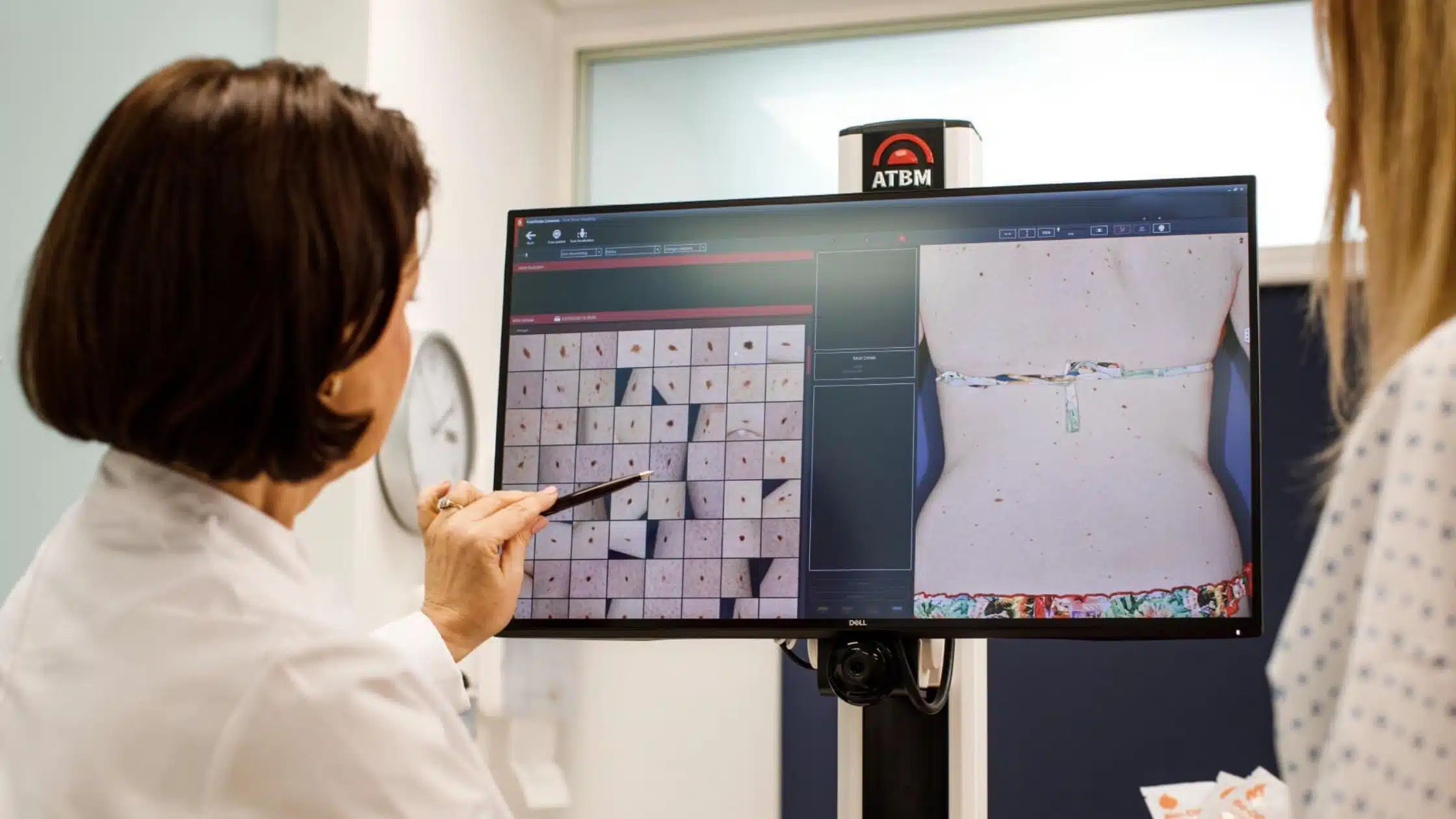
Australia has one of the highest rates of skin cancer in the world, making regular skin checks an important part of staying healthy. In Canberra, where outdoor activities are part of everyday life, prolonged exposure to ultraviolet (UV) radiation can increase the risk of developing skin cancer. While many moles and skin spots are harmless, some can change over time and become cancerous without causing pain or noticeable symptoms.
This is where Digital Mole Mapping in Canberra plays an important role. Digital mole mapping is an advanced technology that helps monitor moles and pigmented skin lesions over time using high-resolution photography and specialised imaging. Instead of relying only on memory or visual comparison, doctors can accurately identify subtle changes that may indicate the early stages of skin cancer.
Whether you have many moles, a family history of melanoma, or simply want peace of mind, digital mole mapping provides an effective way to support early detection. When combined with regular skin cancer screening, it can help identify suspicious changes before they become more serious.
In this guide, you’ll learn how digital mole mapping works, who should consider it, what happens during the appointment, and why it is becoming an important part of skin cancer prevention in Canberra.
Why Early Detection of Skin Cancer Matters

Australia has very high levels of UV throughout much of the year, meaning that even cooler or cloudy days can lead to damage of the skin.
Many skin cancers grow slowly, and if not treated, others — especially melanomas — can grow rapidly. Early detection of potentially abnormal changes can significantly improve treatment, recovery, and long term outcomes.
If caught early, there are a number of important benefits:
- The good news is that higher treatment success rates.
- Treatment options that are less invasive.
- Better cosmetic results.
- Minimized danger of cancer spreading.
- Lower treatment costs.
- More relaxed lifestyle.
The regular skin exam, in addition to digital monitoring, adds another layer of protection, particularly for those with a large number of moles and/or lesions that are hard to monitor.
What is Digital Mole Mapping?
Digital mole mapping is an advanced skin monitoring technique that uses specialised cameras and imaging software to photograph your skin and individual moles.
Mole mapping provides a permanent visual record of your skin, in contrast to a conventional skin examination that is done at one appointment. These images are safely kept and compared to subsequent photographs at your subsequent appointments.
Changes in any of the following, which may not be seen with the naked eye, can be detected by doctors:
- Size
- Shape
- Colour
- Border
- Surface texture
- New mole development
Digital mole mapping facilitates more accurate skin surveillance over time and minimises the removal of non-cancerous moles and ensures that their suspicious ones are attended to.
How Digital Mole Mapping Works
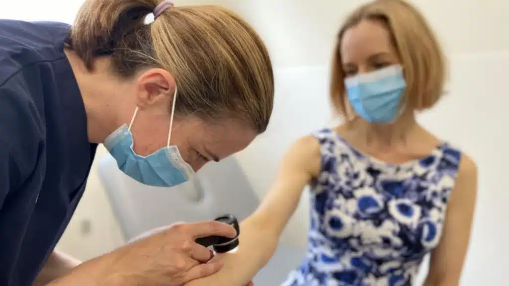
Step 1: To Perform a Complete Skin Examination
The initial portion of your skin cancer doctor appointment will involve a thorough assessment of your skin. Your skin is thoroughly evaluated from head to toe to determine if you need to see something more in-depth.
Step 2: Shot a Photo of Your Entire Body
A series of high resolution photos are made to record the entire surface of your skin.
These images provide a baseline to compare at future appointments.
Step 3: Dermoscopic Imaging
The special dermatoscope is used to photograph the suspect or unusual moles.
This device enlarges the image of skin structures underneath the skin, enabling doctors to check the patterns of pigmentation and various other features that are not visible with the naked eye.
Step 4: Digital Storage
Your pictures are safely kept in your medical record.
These images are compared with photographs taken at future appointments, to identify even small changes.
Step 5: Follow-Up Monitoring
Your doctor might order further imaging tests every six to 12 months depending on your individual risk.
Overtime monitoring will be able to find any changes much sooner than using visual memory.
Who Should Consider Digital Mole Mapping?
Digital mole mapping is useful for anyone but especially those with a higher risk for skin cancer, but regular skin examinations are beneficial for everyone.
You might benefit if you:
- Ensure that there are over 50 moles.
- Have unusual or atypical moles.
- Have previously had melanoma.
- Have had other forms of skin cancer.
- Be a family member of a person with skin cancer melanoma.
- Burn easily and have fair skin.
- Play outside, spending a lot of time outdoors.
- Suffered a number of sunburns.
- Be aware of any new or unusual moles.
- Lack of ability to observe moles on back or head.
For them, frequent photographic surveillance will ensure that any changes will be captured as soon as possible.
Benefits of Digital Mole Mapping
A digital mole map provides a number of benefits over just relying on regular, visual examinations.
Earlier Detection
The biggest benefit would be the fact that you can catch significant changes early before they become apparent.
It can be as simple as a change in colour or shape that can mean early melanoma, meaning treatment can start earlier.
Improved Accuracy
If you compare good pictures taken months or years apart, you’re more likely to produce accurate monitoring than using the power of your own memory.
The doctor can tell if the mole has stayed the same or if it has changed.
Fewer Unnecessary Biopsies
Not all moles with odd appearances need to be dug up.
If past pictures demonstrate that the mole hasn’t altered, unneeded biopsies can also be prevented without endangering the patient’s safety.
Better Long-Term Monitoring
It can be challenging for patients to remember which moles have changed in their bodies when they have numerous moles.
It’s much easier to be able to do long term surveillance with digital records.
Increased Patient Confidence
Understand that the skin is being viewed using cutting-edge imaging and rest assured between visits.
Your doctor and you both have pictures to look back on to determine if a mole has changed or not.
Digital Mole Mapping vs a Standard Skin Check
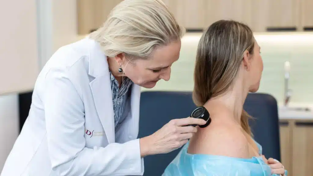
The typical skin cancer examination includes a single appointment during which the skin is checked for abnormalities.
Digital mole mapping takes it one step further and produces a permanent photographic record of the moles, which enables comparisons to be made over months and years.
A regular skin check discovers any abnormal growths that are there now.
One of the first signs of melanoma is the changes identified by digital mole mapping over time.
Digital mole mapping is not a substitute for a skin cancer check, but works in tandem to help monitor and diagnose skin cancer early.
What to Expect During Your Appointment
A digital mole mapping appointment is a relatively simple process, but many people are not aware of what will occur.
Your doctor will ask some questions before your exam, such as asking about your medical history, any other skin cancers you may have had, your family history of melanoma, medications you are taking, and any skin changes you have noticed. Providing this information can be useful to consider your personal risk and the most suitable monitoring plan.
You’ll be asked to undress as necessary during the appointment so that the skin can be examined thoroughly. Then, high-resolution photos of your skin are taken to capture it from several angles. Dermoscopic images are taken with special equipment, which magnifies the image of the skin, if there are moles or lesions that need to be examined further.
Your doctor will talk with you about anything that he or she finds during the exam. Additional work up including biopsy or removal may be suggested if a suspicious lesion is found. If there are no changes of concern, your images are your reference to make in the future.
The appointment usually takes 30-60 minutes, depending on the number of moles that are being assessed and the need for further imaging.
How Often Should You Have Digital Mole Mapping?
The optimal rate is based on individual risk factors and your doctor’s advice.
Some only need to be monitored once a year, others with a greater risk of developing melanoma may need to be monitored more often.
If you are advised to have regular check-ups you:
- Had a previous diagnosis of melanoma in the past.
- Possess a lot of moles.
- Create new moles often.
- Have moles that are different from others.
- I have a family history of skin cancer.
- Stay outdoors for a long period of time because of work or recreation.
For patients, repeat imaging tests are useful at 6-12 month intervals for patients, a balance between continued surveillance and early detection.
Signs That Should Never Be Ignored
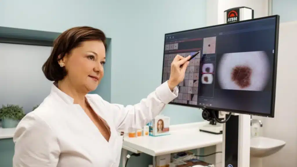
If you have regular skin checks it is still important to keep an eye out for changes between check-ups.
If you see any of the following, arrange a skin assessment immediately:
- A new mole that has emerged when adult.
- A mole that has a different size, shape or color.
- Sides that are not straight or even.
- A mole with several different colours in it.
- A sore that does not heal.
- Rough, crusty, itchy skin spots.
- An area that seems to be different from other moles.
ABCDE Guide
A – Asymmetry
One half of the mole is different from the other.
B – Border
The edges are indistinct, irregular, or fuzzy.
C – Colour
There are different shades of brown, black, red, white or blue within the same lesion.
D – Diameter
The mole is larger than about 6 mm or it is growing.
E – Evolving
The mole’s appearance/manifestations are changing.
These symptoms are not always a sign of skin cancer, but should always be evaluated by a health care provider.
Why Digital Mole Mapping Is Valuable in Canberra
Canberra residents are likely to be active outdoors, enjoy gardening, walking, cycling, hiking and other outdoor recreation activities that may involve increased exposure to UV radiation. During the colder seasons, the UV levels can still be high enough to cause skin damage.
As sun damage is cumulative, continuous monitoring is crucial for individuals of all ages, especially those with fair skin or a history of heavy sun exposure.
Digital mole mapping will give a further level of confidence as it will document your skin over time. Doctors are not only able to review images in their mind, but also to compare exact images and see small differences that might not have been noticed otherwise.
This strategy helps to identify skin cancer as early as possible, and may help with earlier detection and more successful treatment if it does occur.
Tips for Protecting Your Skin
Digital mole mapping is just one aspect of healthy skin. Regular monitoring and good sun protection measures can help minimise the likelihood of future damage.
To protect the skin you can do the following:
- Use a multi-spectrum Sun Protection Factor (SPF) 50+ sunscreen daily.
- Reapply sun cream every 2 hours when exposed to the sun.
- Use protective clothes, a wide-brimmed hat and UV-protective sunglasses.
- Avoid being in the sun in the middle of the day when it is the hottest and sunniest.
- Don’t over-expose skin to the sun or tanning.
- Regularly check their skin, get to know their moles.
- Have regular professional skin cancer checks based on your personal risk.
These are just a few small tips that will have a huge impact over time.
Frequently Asked Questions
Q1. What is digital mole mapping?
Digital mole mapping is an advanced skin monitoring technique that uses high-resolution photography and dermoscopic imaging to record and track moles over time. By comparing images from different appointments, doctors can detect subtle changes that may indicate the early development of skin cancer.
Q2. Is digital mole mapping painful?
No. The procedure is completely non-invasive and painless. It involves taking photographs of your skin and examining selected moles with specialised imaging equipment. No needles or surgical procedures are involved unless a suspicious lesion requires a biopsy.
Q3. Who should have digital mole mapping?
Digital mole mapping is especially beneficial for people with many moles, atypical moles, fair skin, a personal or family history of melanoma, or those with significant sun exposure. Your doctor can recommend whether this monitoring approach is suitable for your individual risk.
Q4. How often should I have mole mapping?
The recommended frequency varies depending on your risk factors. Many people benefit from annual monitoring, while those at higher risk may be advised to return every six months. Your doctor will recommend a schedule based on your skin health and medical history.
Q5. Does digital mole mapping replace a skin cancer check?
No. Digital mole mapping complements a professional skin examination rather than replacing it. A skin cancer check allows your doctor to assess your skin during the appointment, while mole mapping provides long-term photographic comparisons to detect changes that may occur over time.
Book Your Skin Assessment at Canberra Skin Cancer Clinic
Protecting your skin starts with regular monitoring and early detection. At Canberra Skin Cancer Clinic, our experienced team provides comprehensive skin cancer screening and advanced digital mole mapping to help identify suspicious changes as early as possible.
Whether you need your first skin check or ongoing monitoring of multiple moles, we’re here to support your long-term skin health. Book your appointment today and take a proactive step towards protecting your skin.
| Canberra Skin Cancer Clinic- Green Way | Canberra Skin Cancer Clinic- Molonglo Valley |
| Phone: (02) 51046409 | Phone: (02) 51046409 |
| Email: reception@greenwaymedicalcentre.com.au | Email: reception@greenwaymedicalcentre.com.au |
| Address: Unit 6/175 Anketell Street, Greenway ACT 2900 | Address: 110 Woodberry Ave Coombs ACT 2611 |
| Website: https://canberraskincancerclinic.com.au/ | Website: https://canberraskincancerclinic.com.au/ |