
A new, changing or unusual skin lesion can be difficult to assess on your own. Many harmless moles, freckles, cysts and other skin growths can look similar to skin cancer, while some early skin cancers may appear subtle and cause no pain or other symptoms.
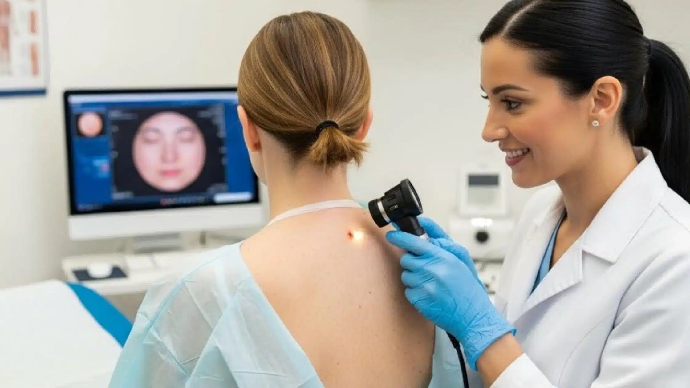
Skin Lesion Assessment in Canberra provides a structured approach to examining suspicious or changing areas of skin. A medical professional may assess the lesion visually, use dermoscopy to examine structures beneath the skin surface, compare the lesion with previous images where appropriate, and recommend a biopsy when further investigation is needed.
Early assessment does not mean that a lesion is cancerous. Instead, it helps determine whether a spot can be safely monitored or requires further investigation.
For Canberra residents, understanding when a lesion should be checked and what to expect during an assessment can make it easier to seek appropriate medical advice.
What Is a Skin Lesion?
A skin lesion is a general term for an area of skin that looks or feels different from the surrounding skin.
Skin lesions can include:
- Moles or naevi
- Freckles
- Pigmented spots
- Cysts
- Seborrhoeic keratoses
- Skin tags
- Actinic keratoses
- Benign growths
- Basal cell carcinomas
- Squamous cell carcinomas
- Melanomas
The term “lesion” does not automatically mean cancer.
Some lesions are completely harmless, while others may require treatment or monitoring. The purpose of a professional assessment is to distinguish between these possibilities as accurately as possible.
Why Skin Lesion Assessment Matters
Skin cancer can sometimes develop without obvious symptoms.
A lesion may not hurt, itch or bleed, particularly during its early stages. This means that waiting for symptoms before arranging an examination is not always appropriate.
Changes in the appearance of a mole or skin lesion can be an important warning sign. A lesion that changes in size, shape or colour should be assessed by a medical professional.
Skin cancer is common in Australia, and ultraviolet (UV) radiation is a major risk factor. Regular awareness of your skin and appropriate professional assessment can help identify suspicious changes earlier.
Skin Lesion Assessment in Canberra: What Happens?
The assessment process depends on the appearance of the lesion and your individual risk factors.
A typical appointment may involve several stages.
1. Medical History
Your clinician may ask questions about the lesion and your general skin cancer risk.
This may include:
- When you first noticed the lesion
- Whether it has changed
- Previous skin cancers
- Previous biopsies
- Family history of melanoma
- Sun exposure
- History of sunburn
- Number and type of moles
- Previous treatment of suspicious lesions
Providing information about when you first noticed a change can be particularly helpful.
2. Visual Examination
The clinician examines the lesion and surrounding skin.
The assessment may consider:
- Shape
- Size
- Border
- Colour
- Surface
- Symmetry
- Texture
- Location
- Changes over time
The surrounding skin may also be assessed because an isolated examination of only the lesion you noticed may not provide the complete clinical picture.
3. Dermoscopy
Dermoscopy is an important part of modern skin lesion assessment.
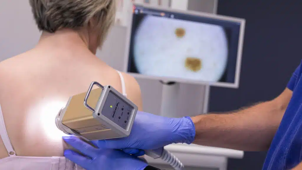
A dermatoscope is a specialised magnifying device that allows trained clinicians to examine pigmentation patterns and structures that are difficult to see with the naked eye.
Dermoscopy can help distinguish lesions that appear suspicious from those that have features more consistent with benign conditions.
However, dermoscopy is not a substitute for pathology when a lesion requires definitive diagnosis.
When Does a Skin Lesion Need Further Investigation?
Not every unusual-looking lesion needs to be removed.
Depending on the clinical findings, a lesion may be:
- Considered benign and require no treatment
- Monitored over time
- Photographed for comparison
- Re-examined at a future appointment
- Biopsied or removed for pathology
The appropriate approach depends on the lesion and the person’s individual risk factors.
A lesion that appears stable and clearly benign may not require intervention. A lesion that is changing or has suspicious dermoscopic features may need further investigation.
Signs That a Skin Lesion Should Be Checked
You should consider arranging a professional assessment if you notice a lesion that is:
- New
- Changing in size
- Changing in shape
- Changing in colour
- Becoming darker
- Developing multiple colours
- Developing an irregular border
- Increasing in thickness
- Persistently crusting
- Bleeding without an obvious cause
- Not healing
- Different from your other moles
- Causing persistent concern
One useful principle is evolution.
A lesion that looks unusual but remains stable may be less concerning than a lesion that has recently changed. However, appearance alone cannot determine whether a lesion is cancerous.
Understanding the ABCDE Approach
The ABCDE method is commonly used as a guide for recognising potentially suspicious pigmented lesions.
A — Asymmetry
One half of the lesion may look different from the other.
B — Border
The edge may appear irregular, uneven or poorly defined.
C — Colour
The lesion may contain different shades or areas of uneven pigmentation.
D — Diameter
Larger lesions can be concerning, although melanoma can also occur in lesions smaller than 6 mm.
E — Evolution
Changes in size, shape, colour or appearance are particularly important.
The ABCDE method is useful for awareness but should not be used as a self-diagnosis tool. Some melanomas do not display all of these features.
Skin Lesions That Can Resemble Skin Cancer
One reason professional assessment is valuable is that benign and malignant lesions can sometimes appear similar.
For example, a harmless seborrhoeic keratosis can look unusual or pigmented. An inflamed cyst may appear red and swollen. An actinic keratosis may appear as a rough or scaly patch.
At the same time, some skin cancers can appear relatively subtle.
This is why a lesion should be assessed according to its clinical characteristics rather than based solely on whether it “looks like cancer”.
Skin Lesion Assessment and Melanoma Detection
Melanoma develops from melanocytes, the pigment-producing cells of the skin.
It can develop within an existing mole or appear as a new lesion.
Some melanoma warning signs include:
- A new pigmented lesion
- A changing mole
- Uneven pigmentation
- An irregular border
- A lesion that looks different from surrounding moles
- A lesion that continues to grow
- A changing lesion that develops symptoms
Melanoma can also occur in less obvious locations, including the scalp, soles of the feet, palms and underneath nails.
For this reason, a complete skin examination may be appropriate for people with relevant risk factors rather than focusing exclusively on the lesion that first attracted attention.
Melanoma Diagnosis Canberra: Why Professional Assessment Is Important
Melanoma diagnosis Canberra services generally involve clinical examination and dermoscopy, followed by biopsy when a lesion is considered suspicious.
It is important to understand that a clinician cannot always confirm melanoma simply by looking at a lesion.
If melanoma is suspected, tissue may need to be removed and examined by a pathologist.
The pathology examination can determine whether melanoma is present and provide information about the characteristics of the tumour.
This information can then guide further management.
Melanoma Screening and High-Risk Patients
There is no single screening schedule that is appropriate for everyone.
People with certain risk factors may benefit from more regular professional skin examinations.
Risk factors can include:
- Previous melanoma
- A family history of melanoma
- Numerous moles
- Atypical naevi
- Significant UV exposure
- Previous skin cancers
- Repeated sunburn
- Fair skin that burns easily
People with many moles may also find it difficult to identify subtle changes themselves.
In selected patients, total body photography or sequential digital dermoscopy may be considered to help identify changes over time.
Melanoma screening should therefore be tailored to individual risk rather than based on a one-size-fits-all schedule.
What Is Digital Monitoring?
Digital monitoring involves taking photographs or dermoscopic images of selected lesions and comparing them during future examinations.
This can be useful when a lesion does not clearly require immediate biopsy but has features that justify observation.
For patients with multiple moles, total body photography can also help clinicians identify new lesions or changes between appointments.
Digital monitoring is not suitable for every lesion. A lesion that appears sufficiently suspicious may need biopsy rather than observation.
When Is a Skin Biopsy Recommended?
A biopsy may be recommended when clinical examination and dermoscopy cannot confidently establish that a lesion is benign.
For a lesion suspected to be melanoma, an excision biopsy is commonly used when practical.
The area is numbed with local anaesthetic before the lesion is removed. The tissue is then sent to a pathology laboratory for microscopic examination.
The pathology report provides the definitive diagnosis and may describe features that are important for determining the next stage of care.
Skin Cancer Diagnosis: Why Pathology Matters
Skin cancer diagnosis cannot always be established from appearance alone.
Different skin cancers can have different clinical appearances, and benign lesions can sometimes mimic malignancy.
Pathology allows a specialist pathologist to examine the cells and tissue architecture under a microscope.
Depending on the lesion, pathology can determine whether it represents:
- Benign tissue
- Precancerous change
- Basal cell carcinoma
- Squamous cell carcinoma
- Melanoma
- Another skin condition
If cancer is identified, the report may also contain information that helps determine appropriate treatment.
Common Types of Skin Cancer
Basal Cell Carcinoma
Basal cell carcinoma is the most common type of skin cancer.
It often develops on areas of skin that receive substantial UV exposure, such as the face, ears, scalp and neck.
It may appear as a pearly lump, persistent sore, scaly area or lesion that repeatedly bleeds or crusts.
Squamous Cell Carcinoma
Squamous cell carcinoma can develop on sun-damaged skin.
It may appear as a scaly or crusted patch, a firm lump or a sore that does not heal.
Some SCCs can grow more aggressively than others, making timely assessment important.
Melanoma
Melanoma develops from melanocytes and can potentially spread if it becomes invasive.
A new or changing pigmented lesion should be assessed rather than assumed to be harmless.
Skin Lesion Assessment for Canberra Residents

Canberra residents may spend time outdoors for work, sport, recreation, gardening, cycling, walking and other activities.
UV exposure accumulates over time, and sun damage can contribute to the development of skin cancers.
This makes skin awareness particularly relevant for people who regularly spend time outdoors.
However, skin cancer can affect people who do not consider themselves to be high risk.
A professional assessment should therefore be based on your skin, medical history and risk factors rather than assumptions about whether you have had “enough” sun exposure to be concerned.
Can You Diagnose a Skin Lesion at Home?
It is useful to monitor your skin at home, but self-examination cannot provide a definitive diagnosis.
You can look for changes in:
- Size
- Shape
- Colour
- Texture
- Thickness
- Border
- Symptoms
Photographs taken over time can also help you remember how a lesion looked previously.
However, if a lesion appears suspicious or changes noticeably, professional assessment is recommended.
Online image searches and smartphone applications should not be relied upon to determine whether a lesion is cancerous.
How Often Should Skin Lesions Be Checked?
The appropriate frequency depends on your individual circumstances.
Someone with no significant risk factors and no suspicious lesions may not require the same monitoring schedule as someone with multiple atypical moles or a previous melanoma.
Your clinician can recommend an appropriate follow-up interval based on your risk profile and examination findings.
Importantly, you should arrange an assessment when you notice a significant new change rather than waiting for a scheduled appointment.
How to Prepare for Your Skin Lesion Assessment
Before your appointment, consider making a note of:
- When you first noticed the lesion
- Any changes you have observed
- Whether it has bled or crusted
- Previous skin cancer diagnoses
- Family history of melanoma
- Previous biopsies
- Medications or relevant medical information
If you have photographs showing how the lesion looked previously, these may also be useful.
Try to identify any other lesions that have recently changed so you can mention them during the consultation.
What Happens If the Lesion Is Benign?
A benign result can be reassuring.
Depending on the type of lesion, no treatment may be necessary.
Some benign lesions can be removed if they are repeatedly irritated, symptomatic or otherwise troublesome, although removal is not always medically necessary.
Your clinician can explain whether the lesion needs monitoring and whether you should return for a future skin examination.
What Happens If Skin Cancer Is Found?
If pathology confirms skin cancer, your clinician can discuss the diagnosis and treatment options.
Treatment depends on the type of skin cancer, its location, size and other clinical factors.
Possible treatments can include:
- Surgical excision
- Curettage and cautery for selected lesions
- Cryotherapy for appropriate precancerous lesions
- Topical treatments for selected conditions
- Specialist treatments for more complex cancers
Melanoma treatment is determined according to the stage and pathological characteristics.
A confirmed diagnosis should therefore be followed by an individualised treatment plan rather than a generic approach.
Melanoma Detection: Why Changes Matter
Melanoma detection is not simply about identifying a large or very dark mole.
Some melanomas may be small, while others may not have a classic appearance.
A lesion that is changing or looks significantly different from the rest of your moles can deserve assessment even when it does not fit every ABCDE warning sign.
The most useful approach is to become familiar with your skin and seek professional advice when something changes.
Preventing Further Skin Damage
Skin lesion assessment is only one part of skin cancer prevention.
Reducing unnecessary UV exposure can help protect the skin.
Sun protection measures include:
- Using broad-spectrum sunscreen appropriately
- Wearing protective clothing
- Wearing a broad-brimmed hat
- Using sunglasses
- Seeking shade
- Avoiding unnecessary exposure during periods of high UV
- Reapplying sunscreen as directed
Sun protection should be part of everyday outdoor habits, particularly in Australia.
Why Early Assessment Is Better Than Waiting
A suspicious lesion does not necessarily mean cancer.
However, delaying assessment can mean that a lesion continues to develop without appropriate evaluation.
If a lesion is benign, assessment can provide reassurance. If it is precancerous or cancerous, early identification may allow appropriate treatment to begin sooner.
This is why a changing lesion is worth discussing with a medical professional rather than monitoring indefinitely without guidance.
When to Book a Skin Lesion Assessment in Canberra
Consider arranging an appointment if you have:
- A new or unusual mole
- A changing pigmented lesion
- A spot that looks different from your other moles
- A lesion that continues to grow
- A persistent scaly or crusted patch
- A sore that does not heal
- A lesion that repeatedly bleeds
- A suspicious spot identified during self-examination
- A personal history of skin cancer
- Multiple moles requiring professional monitoring
You do not need to know whether the lesion is melanoma before booking an assessment.
That is the purpose of professional examination.
Frequently Asked Questions
What is included in a skin lesion assessment?
A skin lesion assessment may include a medical history, visual examination and dermoscopy. If a lesion appears suspicious, the clinician may recommend monitoring, photography or biopsy. The exact assessment depends on the appearance of the lesion and the patient’s individual risk factors.
Can dermoscopy diagnose skin cancer?
Dermoscopy can provide valuable information about structures and patterns within a skin lesion that are not easily visible to the naked eye. It can help a trained clinician decide whether a lesion appears benign or suspicious. However, a biopsy and pathology examination may be required for definitive diagnosis.
Should I get a changing mole checked?
Yes. A mole that changes in size, shape or colour should be assessed by a medical professional. Changes do not automatically mean melanoma, but they can be an important warning sign that further evaluation is appropriate.
Can melanoma be detected early?
Yes. Melanoma can sometimes be identified while it is still confined to the skin. Regular awareness of your skin, professional examination when appropriate and prompt assessment of suspicious changes can support earlier detection.
Does every skin lesion need to be removed?
No. Many skin lesions are benign and do not require removal. Depending on its appearance and risk, a lesion may simply be left alone, monitored or photographed. Removal or biopsy may be recommended when the lesion cannot be confidently classified as benign.
What should I do if a skin lesion starts bleeding?
A lesion that repeatedly bleeds, crusts or fails to heal should be assessed by a medical professional. These symptoms can have several causes, but persistent bleeding or non-healing should not be ignored.
Take Suspicious Skin Changes Seriously
Most skin lesions are not cancerous, but it is not always possible to tell the difference between a harmless lesion and an early skin cancer by looking at it yourself.
Skin Lesion Assessment in Canberra provides an opportunity to have new, changing or unusual lesions professionally examined. Clinical assessment, dermoscopy, appropriate monitoring and pathology can each contribute to accurate diagnosis.
If you have noticed a skin lesion that is new, changing, bleeding, growing or simply concerning, arranging a professional assessment is a sensible next step.
Book Your Skin Lesion Assessment in Canberra
If you are concerned about a mole, spot or changing skin lesion, contact Canberra Skin Cancer Clinic to arrange an assessment and discuss whether further investigation is appropriate.