
Melanoma is one of the most serious forms of skin cancer because it can grow deeper into the skin and spread to other parts of the body if it is not detected and treated early. In Australia, where ultraviolet (UV) exposure is a significant risk factor, recognising suspicious skin changes and arranging appropriate medical assessment is particularly important.
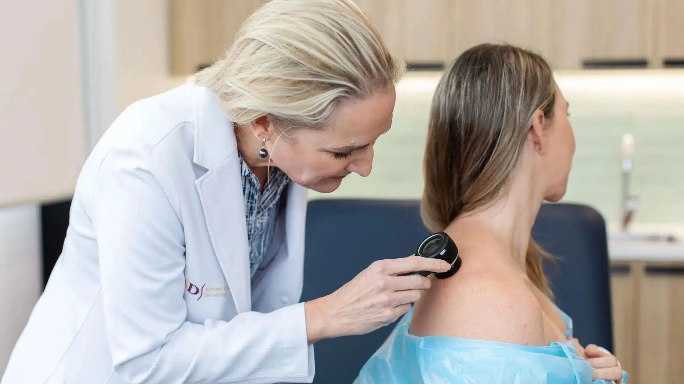
Melanoma Diagnosis in Canberra involves more than simply looking at a mole. A medical professional may assess your skin using clinical examination and dermoscopy, compare suspicious lesions with previous images when appropriate, and recommend a biopsy when melanoma cannot be ruled out. The tissue removed during a biopsy is examined by a pathologist, providing the definitive diagnosis.
For Canberra residents, knowing what to look for and understanding the diagnostic process can make it easier to seek medical attention when a concerning spot appears.
What Is Melanoma?
Melanoma is a type of skin cancer that develops from melanocytes, the cells responsible for producing melanin, the pigment that gives skin its colour. Melanoma most commonly develops on sun-exposed skin, but it can also occur in areas that receive little or no sun exposure.
Melanoma is considered more serious than many other common skin cancers because it has the potential to invade deeper tissues and spread to other parts of the body.
However, early detection can make an important difference. Cancer Council Australia notes that the depth of melanoma invasion is strongly associated with the risk of spread, which is why identifying melanoma while it is still thin is important.
Why Early Melanoma Diagnosis Matters
Melanoma can sometimes look like an ordinary mole, freckle or pigmented spot. This can make it difficult to recognise without appropriate clinical assessment.
A melanoma may begin as a new lesion or develop within an existing mole. Changes in a lesion can be more important than its initial appearance.
Early diagnosis may allow melanoma to be treated before it becomes more invasive. Cancer Council Australia states that early, localised melanoma is generally managed primarily with surgery, and early detection is associated with better outcomes.
This does not mean every changing mole is melanoma. Many skin lesions are benign. The purpose of a professional skin examination is to determine which lesions require closer assessment or further investigation.
Signs That May Require Melanoma Assessment
A useful way to remember some warning signs is the ABCDE approach.
Asymmetry
One half of a mole or pigmented lesion may look different from the other half.
Border
The border may appear irregular, uneven, scalloped or poorly defined.
Colour
A suspicious lesion may contain several shades or areas of different pigmentation rather than having a uniform colour.
Diameter
A lesion larger than approximately 6 mm may warrant assessment, although melanoma can also be smaller than this size.
Evolution
Changes over time are particularly important. A mole that changes in size, shape or colour, or a new lesion that continues to develop, should be assessed by a medical professional.
Cancer Council Australia also notes that some melanomas, including nodular and desmoplastic melanomas, may not follow the typical ABCD pattern.
For this reason, the ABCDE rule should be treated as a guide rather than a diagnostic test.
Other Changes You Should Not Ignore
Not every melanoma appears as a classic dark or irregular mole.
You should consider seeking medical assessment if you notice:
- A new spot that looks different from your other moles
- A mole that is changing in appearance
- A lesion that becomes darker or develops multiple colours
- A spot that continues to grow
- A lesion that becomes raised
- Persistent bleeding or crusting
- An unusual mark that does not settle
- A new pigmented lesion that looks significantly different from surrounding spots
- A changing lesion on the scalp, face, hands, feet or other less obvious areas
Melanoma can also occur in less sun-exposed areas, including the palms, soles and underneath the nails.
Melanoma Diagnosis in Canberra: What Happens During an Examination?
A melanoma assessment generally begins with a detailed examination of the skin.
Your doctor may ask about:
- When you first noticed the lesion
- Whether it has changed
- Previous skin cancers
- Your history of sun exposure
- Your family history of melanoma
- The number and appearance of your moles
- Previous biopsies or skin procedures
The suspicious lesion can then be examined more closely.
Clinical Skin Examination
During a skin examination, the doctor assesses the appearance of moles, freckles, pigmented lesions and other areas of concern.
Depending on your circumstances, a broader examination of the skin may also be appropriate rather than focusing on only the spot you noticed.
This is important because patients may notice one particular lesion while another area of the skin may contain a lesion that requires assessment.
Dermoscopy
Dermoscopy is an important tool used during the assessment of suspicious skin lesions.
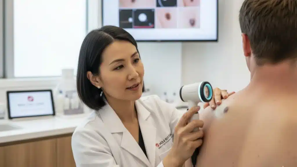
A dermatoscope is a handheld magnifying device that allows a trained clinician to examine structures and pigmentation patterns that may not be visible with the naked eye.
Cancer Council Australia recommends dermoscopy as an aid to clinical diagnosis and notes that its effectiveness depends on appropriate clinician training and experience.
Dermoscopy does not by itself provide a definitive diagnosis of melanoma. Instead, it helps the clinician decide whether a lesion appears benign, requires monitoring or should be removed for pathology.
Digital Monitoring and Mole Mapping
Some people have numerous moles or other factors that place them at higher risk of melanoma. In these situations, photographic monitoring can be useful.
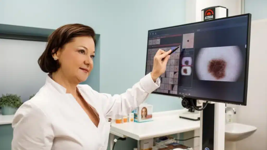
Total body photography creates images that can help clinicians identify changes between examinations. Cancer Council Australia identifies total body photography as an additional aid, particularly for people at higher risk of melanoma or those with a high number of naevi.
Sequential digital dermoscopic imaging may also be used for selected lesions.
This approach can be particularly useful when a lesion does not clearly show melanoma but has characteristics that warrant monitoring.
Importantly, not everyone requires digital monitoring or total body photography. The appropriate approach depends on individual risk factors and clinical findings.
When Is a Biopsy Needed?
Dermoscopy and clinical examination can help identify suspicious lesions, but melanoma cannot always be confirmed by appearance alone.
If melanoma is suspected, the lesion may need to be removed and sent to a pathology laboratory.
For suspected melanoma, an excision biopsy is generally preferred because removing the entire lesion allows the pathologist to examine it more completely. Cancer Council Australia explains that the tissue is examined under a microscope to determine whether melanoma cells are present and to assess important features such as tumour thickness.
The procedure is usually performed using local anaesthetic.
What Happens During an Excision Biopsy?
The area around the lesion is numbed with local anaesthetic.
The doctor then removes the suspicious lesion along with a small amount of surrounding tissue. The wound may be closed with stitches and covered with a dressing.
The specimen is sent to a pathology laboratory.
A pathologist examines the tissue under a microscope and provides a pathology report.
The pathology report is essential because visual examination alone cannot establish the definitive diagnosis.
Understanding the Pathology Report
If melanoma is identified, the pathology report provides important information that helps determine the next steps.
One of the key measurements is Breslow thickness, which describes how deeply the melanoma has invaded the skin.
Other features may also be reported, including ulceration and other microscopic characteristics relevant to staging and treatment.
The information from pathology helps the treating medical team determine whether additional investigation or treatment is required.
Cancer Council Australia explains that melanoma staging ranges from Stage 0 through Stage 4, depending on how far the melanoma has progressed.
What Happens If Melanoma Is Confirmed?
A melanoma diagnosis does not automatically mean that the cancer has spread.
Many melanomas are diagnosed while they are still localised to the skin.
If melanoma is confirmed, your doctor will discuss the pathology results and determine whether further treatment or investigation is appropriate.
For many early melanomas, additional surgery called a wide local excision is performed to remove an appropriate margin of tissue around the original melanoma.
Depending on the characteristics of the melanoma, further investigations may sometimes be recommended.
These can include:
- Ultrasound
- Lymph node assessment
- Sentinel lymph node biopsy
- CT scanning
- MRI
- PET-CT
- Additional pathology or genomic testing
Not everyone with melanoma needs all of these tests. The decision depends on the melanoma’s characteristics and stage.
Melanoma Staging Explained
Staging describes how far melanoma has progressed.
Stage 0
The melanoma is confined to the outermost layer of the skin.
Stage 1
The melanoma remains localised to the original site and has relatively limited thickness.
Stage 2
The melanoma remains localised but has greater thickness and/or other higher-risk characteristics.
Stage 3
The melanoma has spread to nearby lymph nodes or surrounding tissues.
Stage 4
The melanoma has spread to distant areas of the body.
Cancer Council Australia notes that staging is based on pathology findings and, where appropriate, additional investigations.
The stage helps doctors determine the most appropriate treatment pathway.
Who May Be at Higher Risk of Melanoma?
Melanoma can affect anyone, but certain factors can increase risk.
These may include:
- A personal history of melanoma
- A family history of melanoma
- Having many moles
- Having atypical or dysplastic naevi
- Significant UV exposure
- Repeated sunburn
- Outdoor work or recreational activities
- Fair skin that burns easily
- Previous skin cancers
- A history of intense sun exposure during childhood or adulthood
Having a risk factor does not mean that a person will develop melanoma. Likewise, someone without obvious risk factors can still develop melanoma.
This is why paying attention to changes in your skin remains important.
Can Melanoma Occur in Canberra Despite Cooler Weather?
Yes.
UV exposure is not determined simply by how hot the weather feels.
Canberra experiences substantial seasonal variation, and people may spend time outdoors for walking, cycling, gardening, sport and other recreational activities.
UV radiation can damage the skin even when temperatures are comfortable or the day does not feel particularly hot.
Australia has a high incidence of melanoma, and Cancer Council Australia attributes the country’s high melanoma burden partly to substantial solar UV exposure combined with population risk factors.
Why You Should Not Rely Only on Smartphone Apps
There are many smartphone applications marketed as tools for analysing moles or identifying possible skin cancer.
However, these should not replace professional skin assessment.
Cancer Council Australia states that consumer skin cancer apps can be inaccurate and does not recommend using them as a substitute for examination by a doctor.
If you have a changing or suspicious lesion, an in-person medical assessment is a safer approach.
Melanoma Diagnosis in Canberra for People With Multiple Moles
People with numerous moles may find it difficult to identify subtle changes themselves.
This is one reason professional skin examinations can be valuable for people at increased risk.
During an assessment, a clinician can evaluate individual lesions and determine whether photographic or digital monitoring may be appropriate.
For selected higher-risk patients, monitoring over time can help identify lesions that change in a way that warrants further investigation.
The goal is not necessarily to remove every unusual-looking mole. Instead, appropriate monitoring and targeted biopsy can help distinguish lesions that require treatment from those that can safely be observed.
How Often Should You Have Your Skin Checked?
There is no single skin-check schedule that applies to every person.
Your appropriate examination frequency depends on factors such as:
- Personal history of skin cancer
- Family history
- Number of moles
- Atypical naevi
- Previous melanoma
- UV exposure
- Age
- Individual risk profile
- Changes noticed during self-examination
People at increased risk may require more regular professional assessment.
If you notice a new or changing lesion, you should not wait for your next routine appointment. Arrange an assessment promptly.
Cancer Council Australia encourages people to become familiar with their skin and consult a doctor when they notice changes in the size, shape or colour of a lesion or develop a new lesion.
What You Can Do Between Skin Checks
Professional examinations are only one part of melanoma awareness.
You can also become familiar with your own skin.
When checking your skin, look at areas that are easy to see as well as areas that may be overlooked, including:
- Scalp
- Behind the ears
- Neck
- Back
- Shoulders
- Chest
- Arms
- Legs
- Hands
- Feet
- Soles
- Between the toes
- Under the nails
A partner or family member can help check areas that are difficult to see.
The important point is to look for change.
If something looks different from your other lesions or develops noticeably over time, seek professional advice.
Melanoma Diagnosis in Canberra: When Should You Make an Appointment?
You should consider arranging a skin assessment if you notice:
- A new mole or pigmented lesion
- A mole that is changing
- An irregular border
- Multiple colours within one lesion
- A lesion that is growing
- A spot that looks noticeably different from your other moles
- Persistent bleeding or crusting
- An unusual lesion that concerns you
- A suspicious spot identified during a self-check
You do not need to determine whether a lesion is melanoma yourself.
The purpose of an examination is to have a trained medical professional assess the lesion and determine whether further investigation is required.
Choosing a Skin Cancer Clinic in Canberra
When arranging an assessment, consider whether the clinic provides a comprehensive approach to skin cancer detection.
Useful questions to ask include:
- Does the clinician perform a detailed skin examination?
- Is dermoscopy used when assessing suspicious lesions?
- Can suspicious lesions be biopsied when clinically appropriate?
- Is pathology arranged for biopsied tissue?
- Can higher-risk patients be considered for digital monitoring?
- Will the clinician explain the findings clearly?
- What follow-up is recommended if a lesion requires monitoring?
The right approach should be based on your individual risk and clinical findings rather than simply choosing the shortest appointment available.
Frequently Asked Questions About Melanoma Diagnosis in Canberra
What is the most accurate way to diagnose melanoma?
A clinical examination and dermoscopy can identify lesions that appear suspicious, but a definitive melanoma diagnosis generally requires examination of tissue by a pathologist. When melanoma is suspected, an excision biopsy is commonly preferred so the entire lesion can be assessed.
Can a dermatologist or skin cancer doctor tell if a mole is melanoma?
A trained clinician can assess a mole using clinical examination and dermoscopy and determine whether it appears benign, suspicious or requires removal. However, visual assessment cannot always confirm melanoma. Pathological examination of a biopsy is used to establish the definitive diagnosis.
Can melanoma be smaller than 6 mm?
Yes. Although the ABCDE approach includes diameter greater than approximately 6 mm as one warning sign, melanoma can be smaller. A lesion that is changing, particularly in size, shape or colour, should be assessed regardless of its diameter.
Does every suspicious mole need a biopsy?
No. Some lesions can be assessed clinically and with dermoscopy and may not require immediate removal. Others may need monitoring or biopsy because their appearance or behaviour raises concern. The decision should be made by a qualified medical professional based on the individual lesion.
How long does a melanoma biopsy take?
The biopsy itself is generally a relatively quick procedure performed under local anaesthetic. The tissue is then sent to a pathology laboratory for microscopic examination. The exact timeframe for receiving results can vary between laboratories and clinical circumstances.
What happens after melanoma is diagnosed?
The next step depends on the pathology findings, including melanoma thickness and other features. Many early melanomas require further surgical removal of tissue around the original site. Some patients may need additional lymph node assessment or imaging depending on their stage and risk characteristics.
Is melanoma always caused by sun exposure?
UV radiation is an important risk factor for many melanomas, but melanoma can also develop in areas that receive little sun exposure, including the palms, soles and underneath nails.
Take Changes in Your Skin Seriously
Melanoma can sometimes be difficult to recognise because it may initially resemble an ordinary mole or pigmented spot. A new lesion or a noticeable change in an existing lesion should not automatically be assumed to be harmless.
Melanoma Diagnosis in Canberra starts with recognising when a lesion needs professional attention. Clinical examination, dermoscopy, appropriate monitoring and biopsy can each play an important role in the diagnostic process.
If you have noticed a changing mole, a new unusual spot or another skin lesion that concerns you, consider arranging an assessment rather than trying to diagnose it yourself.
Book a Skin Assessment in Canberra
If you are concerned about a mole or changing skin lesion, Canberra Skin Cancer Clinic can provide professional assessment and advice about whether further investigation is appropriate.
Book an appointment with Canberra Skin Cancer Clinic to have a suspicious or changing skin lesion assessed by a qualified medical professional.