Solar Keratoses (Sun Spots) in Canberra are a common sign of long-term ultraviolet (UV) damage. Also known as actinic keratoses or solar keratoses, these areas can appear as rough, dry, scaly or thickened patches on skin that has received repeated sun exposure.
Solar keratoses are not the same as invasive skin cancer. However, they are an important marker of sun-damaged skin and can sometimes develop into squamous cell carcinoma (SCC). People with multiple solar keratoses may also have an increased overall Skin Cancer Risk, particularly when combined with other risk factors such as significant UV exposure, fair skin or a history of skin cancer.
Understanding how these lesions are diagnosed, what changes may require attention and how sun damage relates to skin cancer can help Canberra residents make informed decisions about their skin health.
What Are Solar Keratoses?
Solar keratoses occur as an abnormal growth of skin, primarily due to long-term exposure to UV rays. As time passes, the ultraviolet radiation can damage the skin cells and cause changes to the outer skin cells.
They are more common on areas of the body that are exposed to sun frequently, including the face, ears, scalp, neck, hands, forearms and lower legs. Sometimes a lesion is easier to feel than see (particularly if it is small). It may be rough or gritty like fine sandpaper.
This depends on the individual and can look different for each person. The colour of a solar keratosis can range from skin tone to pink to red to white to yellowish or pigmented. Some are flat, and some raised, thickened or crusted. Large areas with sun damage can have several different lesions.
Sun spot may sometimes be confusing as harmless pigmentation is also named as such. Solar keratoses are distinct in that they are often rough or scaly.
Why Do Solar Keratoses Develop?
The main cause is repeated exposure to UV radiation. Sun damage is cumulative, and the damage that occurs at one time in life can affect the skin condition at another time.
Factors that will make you more likely to get solar keratoses:
- Frequent outdoor activity
- Chronic solar damage due to job.Chronic sun exposure exposure to the sun over the course of a career.
- Previous sunburn
- Easily tanned skin or skin that burns with ease.
- Increasing age
- Ill diagnosed skin cancers on previous skin examinations
- Solar keratoses are the result of multiple previous solar keratoses.
- Some health problems or drugs which lower the body’s defenses
Solar keratoses and a high UV risk are among the factors that Cancer Council Australia sees as contributing to a high risk of developing keratoses.
Cumulative exposure can occur from activities undertaken on a daily basis, including outdoor sport, running, cycling, walking and gardening, by Canberra residents. Sun protection is important all year round, even in warm weather.
What Do Solar Keratoses Look and Feel Like?
Being familiar with the common patterns of skin changes can assist you in knowing that you have a skin change that needs medical evaluation.
Roughness or Gritty Surface (Texture)
One of the most prevalent is a rough texture. Although a small lesion may not appear abnormal at first glance, it might feel differently on the skin when touched.
Dry or Scaly Surface
There may be a dry, flaky or scaly texture to the surface of solar keratoses. It can be light at first, and thicken over time.
Pink or Red Areas
Pink or red lesions are seen in some lesions. These may be flat or slightly raised, and can be more apparent on lighter skin.
Skin-coloured (or pigmented) lesions are lesions that are blue or purple in color.
Solar keratoses don’t always appear red. Others may be very close to the surrounding skin colour or brownish or darker in colour.
Thickening
The lesion can get thicker over time or even feel hard and rough. Any area that is getting thicker, especially when it is painful or growing quickly, should be examined
Tenderness or Bleeding
A large number of solar keratoses are asymptomatic. Others may be tender or sensitive. If these are present, such as if the lesion keeps coming back, gets painful, ulcerates or grows, it should be treated by a medical professional as it can be SCC.
Solar Keratoses and Skin Cancer Risk
The link between solar keratoses and skin cancer is one of the foremost reasons to take them seriously.
Solar keratoses are believed to be precursors to cutaneous squamous cell carcinoma and may develop to this disease. A single solar keratosis is unlikely to develop into a cancer, but many are an indicator of extensive sun damage and have a greater chance of developing skin cancer. According to DermNet, those who have more than one AK are at risk of developing skin cancers, especially SCC.
This does not mean that all solar keratoses will turn into cancer. Some will stay the same and some will not be seen or may be less prominent. If a lesion or change is noted, however, it should not just be written off as a sun spot.
One of the lesions, especially, could turn into:
- Thicker
- Larger
- Painful or tender
- Ulcerated
- Rapidly growing
- Repeatedly bleeding
- Increasingly crusted
These changes may overlap with the clinical features of SCC and examination is important.
Some other skin cancer risks have been established with sun damage:
Solar keratoses are a sign of high UV exposure. This does not only relate to SCC.
Individuals who have had significant sun damage also may develop other forms of skin cancer, such as basal cell or melanoma. The Cancer Council Australia lists exposure to the sun as one of the primary risk factors for skin cancer and says that solar keratoses are associated with sun damage and a risk factor for skin cancer.
That is why it is important to take note of the overall appearance of the skin instead of a specific solar keratosis when multiple ones are present.
Other Skin Cancer Risks Associated With Sun Damage
Solar keratoses indicate that the skin has experienced significant UV exposure. This does not only relate to SCC.
People with substantial sun damage can also develop other types of skin cancer, including basal cell carcinoma and melanoma. Cancer Council Australia identifies UV exposure as a major risk factor for skin cancer and notes that solar keratoses are a marker of sun damage and a risk factor for skin cancer.
This is why the presence of several solar keratoses may be a reason to pay closer attention to overall skin health rather than focusing on one individual spot.
How Are Solar Keratoses Diagnosed?
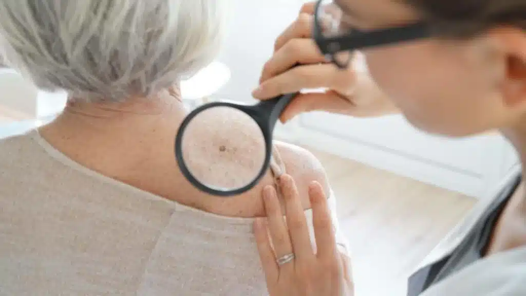
Diagnosis usually begins with a physical examination. A doctor assesses the lesion’s colour, texture, size, thickness, location and overall appearance.
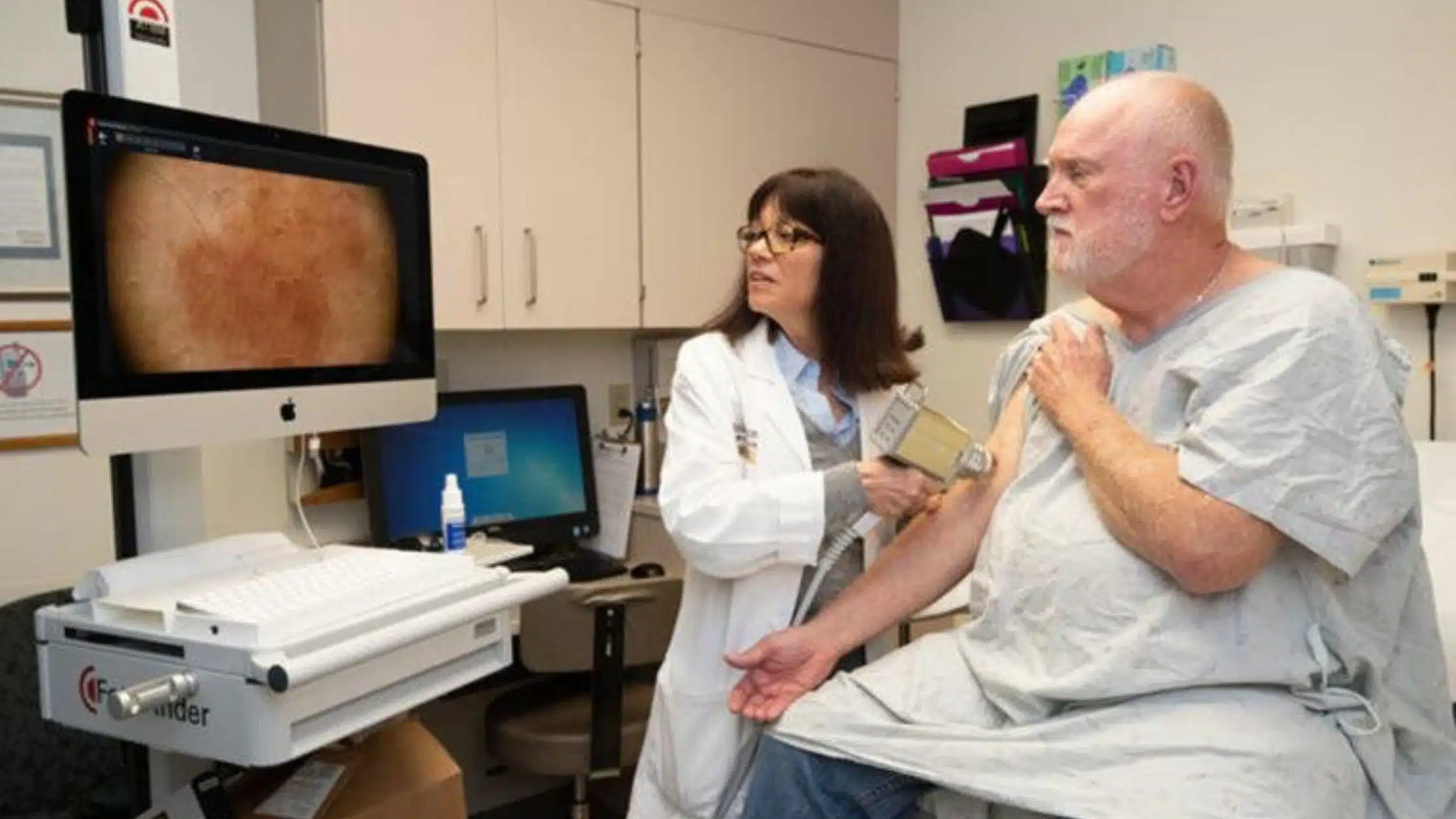
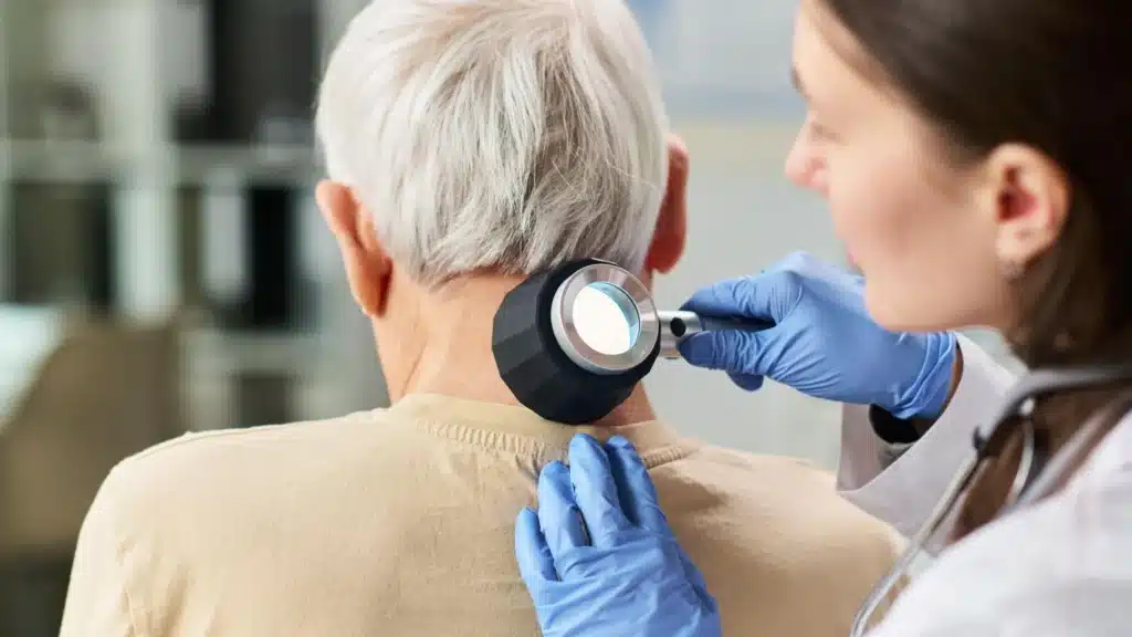
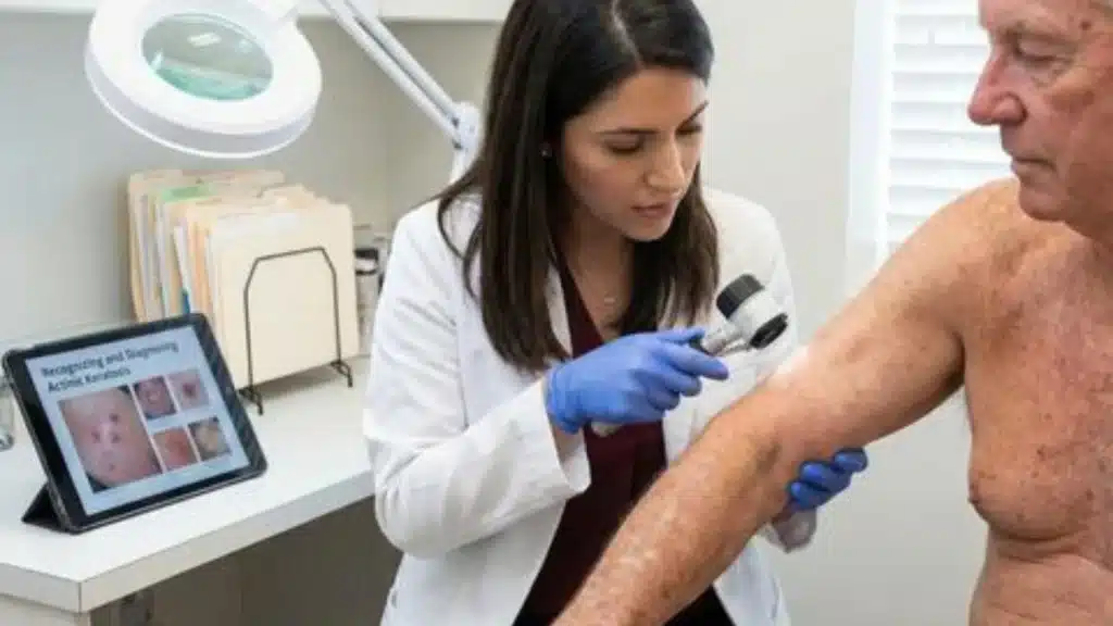
A dermatoscope may also be used. This is a handheld magnifying device that allows a doctor to examine structures within the skin that are not easily visible to the naked eye. Dermoscopy can help distinguish solar keratoses from other lesions, although some lesions can still be difficult to differentiate clinically.
In some cases, a biopsy may be necessary. This can be recommended when:
- The diagnosis is uncertain
- The lesion looks unusually thick
- There is concern about SCC
- The lesion is changing rapidly
- A lesion does not respond as expected to treatment
- The clinical appearance is unusual
A biopsy involves taking a small sample of tissue for examination under a microscope. It can help establish whether the lesion is a solar keratosis or another condition, including skin cancer.
Why Professional Diagnosis Matters
It can be difficult to identify a solar keratosis accurately at home. Rough skin can have many causes, including eczema, psoriasis, benign growths and different forms of skin cancer.
Some early skin cancers may also look relatively subtle. For this reason, a persistent or changing skin lesion should be assessed rather than diagnosed based only on photographs or appearance.
A professional cancer skin check can provide a broader assessment, particularly when a person has several suspicious areas or a history of significant sun exposure.
Cancer Council Australia explains that a skin check involves examining unusual spots and using a dermatoscope to look more closely at lesions.
When Should You Arrange a Cancer Skin Check?
There is no need to wait until a skin change becomes severe before seeking medical advice.
Consider arranging a cancer skin check if you notice a new, persistent or changing area of skin, especially if it is rough, thickened, bleeding or painful.
Professional assessment may also be appropriate for people who have:
- Multiple solar keratoses
- Extensive sun damage
- A history of skin cancer
- Frequent outdoor exposure
- Fair skin that burns easily
- A weakened immune system
- A family or personal history of certain skin cancers
The appropriate frequency of skin examinations depends on individual risk factors. A doctor can discuss whether regular monitoring is suitable for you.
What Happens During a Skin Check?
During a professional examination, a doctor may ask about your personal and family history, previous sun exposure and any skin changes you have noticed.
The skin may then be examined from head to toe, depending on the type of assessment and your individual circumstances.
A dermatoscope can be used to examine suspicious spots more closely. Areas such as the scalp, ears, neck and other difficult-to-see locations may also need attention.
If a lesion requires further investigation, the doctor may recommend a biopsy or another appropriate procedure. Cancer Council Australia notes that doctors may examine both the spots a patient is concerned about and other moles or lesions during a full-body skin check.
When Should You See a Skin Cancer Specialist?
A skin cancer specialist may be involved when a lesion is difficult to diagnose, has concerning features or requires specialist assessment.
Referral or specialist assessment may be considered when there is uncertainty about whether a lesion represents solar keratosis, SCC, basal cell carcinoma or another skin condition.
A specialist assessment can be particularly useful for lesions that are:
- Rapidly changing
- Thick or firm
- Painful
- Ulcerated
- Repeatedly bleeding
- Not healing
- Resistant to previous treatment
This does not mean that every unusual lesion is cancer. It means that certain features warrant closer examination.
Can Solar Keratoses Be Treated?
Yes. Treatment depends on the number and appearance of lesions, their location, thickness and the clinical assessment.
Common treatment options include cryotherapy, prescription creams, curettage and photodynamic therapy. Some individual lesions may require removal or biopsy when there is concern about possible SCC.
The appropriate treatment should be selected by a qualified medical professional after examining the skin.
Treatment can remove existing lesions, but it does not erase previous UV damage. New solar keratoses may therefore develop in the future.
Reducing Future Skin Damage
Sun protection is important for people with existing solar keratoses and for those who want to reduce further UV damage.
Practical measures include:
- Apply broad-spectrum SPF 50+ sunscreen as directed.
- Reapply sunscreen when required.
- Wear a broad-brimmed hat when outdoors.
- Choose protective clothing where practical.
- Use UV-protective sunglasses.
- Seek shade whenever possible.
- Avoid unnecessary exposure during periods of high UV.
- Check your skin regularly.
Regular self-checks can help you become familiar with the normal appearance and texture of your skin. Cancer Council Australia recommends checking for new or changing spots and seeking medical advice if you are concerned about a mark or growth.
Solar Keratoses in Canberra: What Should You Remember?
For people living in Canberra, solar keratoses should be viewed as a sign of cumulative sun damage rather than simply an ordinary cosmetic sun spot.
A rough patch may be harmless, but it may also represent a solar keratosis or another skin condition. If it changes, becomes thicker, bleeds, becomes painful or fails to heal, professional assessment is important.
Having multiple lesions does not mean that skin cancer is present. However, multiple solar keratoses are associated with increased skin cancer risk, making awareness and appropriate monitoring particularly relevant.
Frequently Asked Questions
Q1. Are solar keratoses cancerous?
Solar keratoses are not invasive skin cancer, but they are considered precancerous changes. Some can progress to squamous cell carcinoma. They also indicate significant cumulative sun damage and are associated with increased skin cancer risk.
Q2. Can one solar keratosis become skin cancer?
A single solar keratosis has a relatively low chance of progressing to SCC, but progression is possible. A lesion that becomes thick, tender, ulcerated or enlarged should be assessed promptly.
Q3. Do solar keratoses always need treatment?
Not every lesion is managed in exactly the same way. Treatment decisions depend on the lesion’s appearance, symptoms, location, number and clinical assessment. A doctor can recommend whether treatment, monitoring or further investigation is appropriate.
Q4. Can solar keratoses come back after treatment?
Yes. Treatment can address existing lesions, but previous UV damage remains. New lesions can develop later, which is why ongoing sun protection and skin monitoring remain important.
Q5. Should I have a skin check if I have multiple solar keratoses?
Discuss this with your doctor. Multiple solar keratoses are associated with increased risk of keratinocyte skin cancer, so an assessment can help identify other suspicious lesions and determine an appropriate monitoring plan.
Final Thoughts
Solar Keratoses (Sun Spots) in Canberra can be an important indicator of cumulative UV damage. Although these lesions are not the same as invasive skin cancer, they can sometimes progress to squamous cell carcinoma and are associated with an increased overall Skin Cancer Risk.
Early diagnosis begins with noticing changes in texture, colour, thickness and appearance. A professional examination, including dermoscopy when appropriate, can help distinguish solar keratoses from other skin conditions and identify lesions that may require biopsy or treatment.
If you notice a persistent, thickened, painful, bleeding or changing sun spot, consider arranging a cancer skin check. For people with significant sun damage or multiple suspicious lesions, discussing ongoing monitoring with a skin cancer specialist or doctor can help establish an appropriate approach to skin health.
Book a Skin Cancer Check in Canberra
Noticed a rough, scaly or changing sun spot? Don’t ignore persistent skin changes. Book a professional skin cancer check in Canberra for timely assessment, diagnosis and appropriate care.
| Canberra Skin Cancer Clinic- Green Way | Canberra Skin Cancer Clinic- Molonglo Valley |
| Phone: (02) 51046409 | Phone: (02) 51046409 |
| Email: reception@greenwaymedicalcentre.com.au | Email: reception@greenwaymedicalcentre.com.au |
| Address: Unit 6/175 Anketell Street, Greenway ACT 2900 | Address: 110 Woodberry Ave Coombs ACT 2611 |
| Website: https://canberraskincancerclinic.com.au/ | Website: https://canberraskincancerclinic.com.au/ |